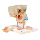
Model Nose With Paranasal Sinuses, illustrates the structure of the nose with the paranasal sinuses in the upper right half of a face in 1.5-fold enlargement, outside of the nose, with paranasal sinuses, differentiated by color, Dimensions : 26 x 19 x 24 cm
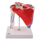
Model Physiology Of Nerves Series, displays the basic structures of the human nervous system, Each of the five sections of the nerve model shows a plastic colored relief model of the main synapse variations, Weight: 4.379 kg, Dimensions : 68 x 51 x 3 cm
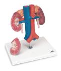
Kidneys with Vessels 2-Part. This kidney model shows the kidneys with suprarenal glands, the outgoing ureters, the renal vessels and the large vessels situated close to the kidneys in natural size.
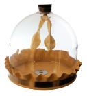
Model Functional Lung Apparatus, unique and easy-to-use model features parts that replicate the actions of respiration, Easy to Assemble, includes an acrylic bell jar, Y tube, two balloons, rubber diaphragm with a knob for ease of movement, and an instruction manual
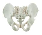
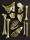
It Illustrate the morphological distinctions between male and female pelvic structures. Each pelvis includes the left and right innominates with pubic symphysis, 4th and 5th lumbar vertebrae with intervertebral discs, the sacrum and coccyx.
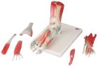
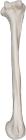
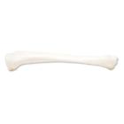
Humerus, right, Model, Human Bone, Plastic, These individual bones are a great addition to you collection, used as a match for your existing Eisco disarticulated skeleton, or to allow students to compare general skeletal structure
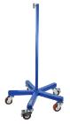
Support base for skeleton, features a heavy (30 pound) cast iron base with 5 machine grade casters (2 with locks) and a sturdy threaded pole, this skeleton stand will pass the test of time and make the transport of y skeleton easy. Inner diameter of stand is 12.5mm.
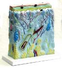
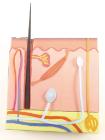
Skin 3-D Model Kit-10 Student Model Template Sets and Teacher Guide, Vinyl Pouch, Assemble and use 3-D models to visualize and investigate key science structures, Perfect for use in the classroom, lab or at home, Satisfies NGSS Standards, Grade: 6 to 10, Material: Paper, LxW: 12X19in
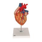
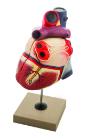
Life size model dissectible in 2 parts. The anterior heart wall can be removed to show the left and right ventricles and atria as well as the tricuspid, pulmonary, mitral and aortic valves. Mounted on base.