Model, Brain, 2 part, Contrasting colors are used to indicate various anatomic structures in the human brain, making this high quality model perfect for beginning anatomy studies, Made of unbreakable vinyl, Dimensions: 15 x 14 x 17.5 cm, weight: 0.82 kg
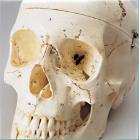
Beauchene Skull Model, 22 Parts, Beauchene, or exploded skull, mounted on stand, Natural color and size, Realistic details, texture and bony landmarks make this model ideal for educational demonstration, model and mount measure 12.75inch in total height
Scapula, right (shoulder blade), Model, Human Bone, Plastic, These individual bones are a great addition to you collection, used as a match for your existing Eisco disarticulated skeleton, or to allow students to compare general skeletal structure
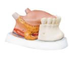
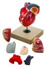
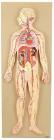
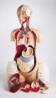
Enlarged. Sectioned so that both ventricles and atria open to expose the valves. Large blood vessels near the heart and musculature of the heart are shown. Separates into 7 parts.
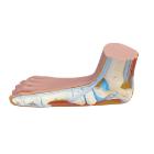
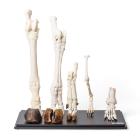
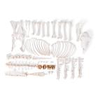
Hind Legs of Different Mammals (Mammalia), Specimen, Included are four real bone specimens: the hind leg of a dog and the hind foot of a horse, a cow and a pig respectively, as well as an original plastic cast of a human foot, Weight: 4 kg
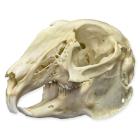
Real Cottontail Rabbit Skull, natural, real bone, The 13 species of Cottontail rabbits can be found throughout much of North America, They are very adaptive and occupy all terrains and most habitats, been mothers for many weeks, Length: 7.5cm (2.9in)
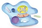
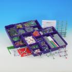
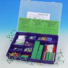
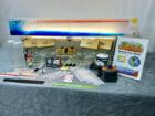
Amoeba 3-D Model Kit-10 Student Model Template Sets and Teacher Guide, Vinyl Pouch, Assemble and use 3-D models to visualize and investigate key science structures, Perfect for use in the classroom, lab or at home, Satisfies NGSS Standards, Grade: 6 to 10, Material: Paper, LxW: 12X19in
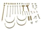
Orbit Hydrogen atom, Hb, Atom centres are colour-coded according to element with prongs set at the correct bonding angles, 10 mm diameter atom centre, Hydrogen bond, Color: White
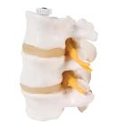
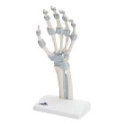
Model, Hand skeleton, with elastic ligaments, The fibrous layer of connective tissue, described in anatomy as the membrana interossea, is shown. It extends between both of these long bones, retinaculum flexorum, Dimensions: 14 x 10 x 28 cm, weight: 0.24 kg
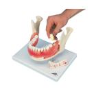
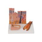
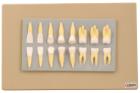
Enlarged approximately 2 times, set of 16 teeth cast in break resistant material having accurate anatomical details. Complete set as in half of upper & lower jaw
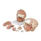
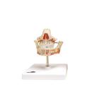
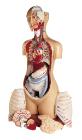
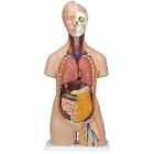
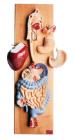
Head and brain 3 part, numbered with english keycard, 3 parts model of the human head which has a removable brain half with arteries, eyeball and optic nerve and the other side exposes the nose, mouth cavity, pharynx etc.
Minit Chlorine atom, Cla, Atom centres are colour-coded according to element with prongs set at the correct bonding angles, 6 mm diameter atom centre, Univalent, Color: Green
Minit Phosphorous atom, Pk, Atom centres are colour-coded according to element with prongs set at the correct bonding angles, 6 mm diameter atom centre, Tetrahedral, Color: Purple
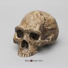
Homo erectus Cranium Model, Designed to give educators affordable, high quality, highly detailed, Durable replica hominins for hands-on study, Lightweight and shatter resistant, Capture fine details, For introductory level courses, Dimension: 20x13.8x12.7cm
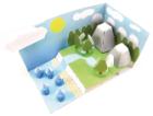
Water Cycle 3-D Model Kit-10 Student Model Template Sets and Teacher Guide, Vinyl Pouch, Assemble and use 3-D models to visualize and investigate key science structures, Perfect for use in the classroom, lab or at home, Satisfies NGSS Standards, Grade: 6 to 10, Material: Paper, LxW: 12X19in
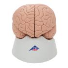
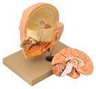
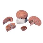
Model Brain 4-Part, right half can be disassembled into: Frontal with parietal lobes, Brain stem with temporal and occipital lobes, Half of cerebellum, great educational tool for the human nervous system and anatomy of the brain, Dimensions : 14 x 14 x 17.5 cm
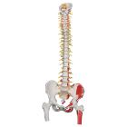
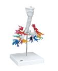
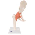
Model Deluxe Functional Hip, high-quality functional hip joint model with ligaments shows the anatomy and possible physiological movements of the human hip joint in exceptional detail, demonstrates abduction, anteversion, retroversion, Dimensions : 32 cm
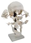
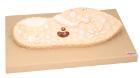
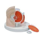
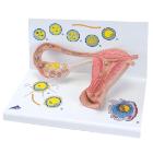
Model Stages Of Fertilisation, Dimensions: 35x21x20 cm, begin the growth into an embryo, The various stages are shown in larger-than-life model form in an ovary, fallopian tube and womb. An even more enlarged illustration of each is also printed on the base