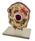
Bio Signs White Blood Cell, Realistic 7-piece model teaches the role of white blood cells and how they keep things moving smoothly through the venous system, Hands-on interactive model provides a magnified and cross-sectioned detailing of a White Blood Cell, With Guide
BioSigns Red Blood Cell Model, Learn all of this and so much more with this hands-on interactive 4-piece model that provides magnified and cross-sectioned detailing of a Red Blood Cell, Develop dexterity, enhance fine motor skills and inspire critical thought
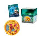
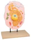
Science BioSigns Animal Cell Model, Hands-on interactive 14-piece model teaches about cell membranes and mitochondria, Provides a magnified and cross-sectioned detailing of an Animal Cell, Includes Guide
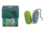
Bacteria Model, Hands-on interactive model provides a magnified and cross-sectioned detailing of Bacteria, Able to be separated, with both halves filled with a cross section of genetic material and flagellum, With Guide
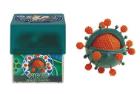
Virus Model, Hands-on interactive model provides a magnified and cross-sectioned detailing of Bacteria, Able to be separated, with both halves filled with a cross section of genetic material and flagellum, With Guide
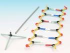
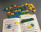
Orbit Small Dna Model Class Kit, Helps students understand DNA, Contains twelve model kits and instructions for guided learning, Students make models of 6 base pairs each that can be joined together to make a molecule of up to 72 base pairs
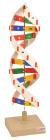
Easy to construct the three dimensional model of DNA. Emphasizing the base pair sequence and function of DNA, the sturdy, colorful bases snap together in the correct sequence, and the pairs attach to a center rod representing hydrogen bonds.
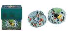
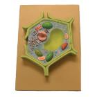
This plant cell model is separated in 4 parts. Showing details of cell wall and inner details of cell wall, nucleus is separated and defined in a 3-D way cut section of chloroplast is shown.
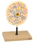
Enlarged 20,000 times. The model shows delicate structure of an animal cell. Features organelles nucleus, endoplasmic reticulum, mitochondria, ribosomes respectively polysomic and Golgi apparatus. Also showing centrioles, lysosomes and fat vacuoles.
Three dimensional model showing electron microscopic structure. Organs like nucleus, Nucleolus, endoplasmic reticulum, mitochondria, ribosomes respectively polysomes and golgi apparatus. Showing centrioles, lysosomes and vacuoles.