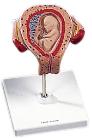
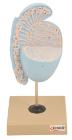
Model, Embryo, 3 month, The embryo model shows an embryo at the stage of the first month of pregnancy, Ideal for medical training and as an educational tool for pregnant women, Important anatomical structures are labeled
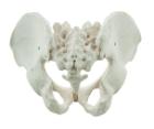
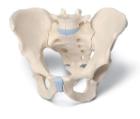
It Illustrate the morphological distinctions between male and female pelvic structures. Each pelvis includes the left and right innominates with pubic symphysis, 4th and 5th lumbar vertebrae with intervertebral discs, the sacrum and coccyx.
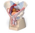
Male Pelvis, 3-Part, This 3-part model is a natural cast of a male, bone pelvis. It shows all anatomical structures in detail: both hip bones, pubic symphisis, sacrum and coccyx as well as the fifth lumbar vertebra with intervertebral disc
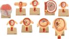
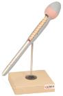
Set of nine models, showing the following stages. 1. Embryo 6 days old 2. 1st month of gestation. 3. Uterus with embryo in 3rd month of gestation.4. Uterus with fetus, in 4th month. 5. Uterus with fetus, placenta and umbilical cord.6. 5th month. 7. 7th month pregnancy.
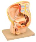
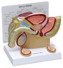
Male Pelvis With Ligmanets, Vessles And Pelvic Floor Organs, This 7 part model of the male pelvis shows in accurate detail how the bones, ligaments, vessels and nerves as well as the pelvic floor muscles and the external sex organs are connected to each other
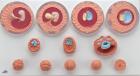
Model, Embryonic Development, 12 stages, represents the development of the human germ cells from fertilisation until the end of the 2nd month of pregnancy in 12 stages, tests for the embryological specialist field, Dimensions: 65 x 34.5 x 6 cm, weight: 0.832 kg