Model, Embryo, 3 month, The embryo model shows an embryo at the stage of the first month of pregnancy, Ideal for medical training and as an educational tool for pregnant women, Important anatomical structures are labeled
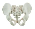
It Illustrate the morphological distinctions between male and female pelvic structures. Each pelvis includes the left and right innominates with pubic symphysis, 4th and 5th lumbar vertebrae with intervertebral discs, the sacrum and coccyx.
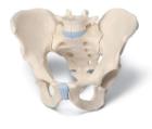
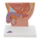
Male Pelvis, 3-Part, This 3-part model is a natural cast of a male, bone pelvis. It shows all anatomical structures in detail: both hip bones, pubic symphisis, sacrum and coccyx as well as the fifth lumbar vertebra with intervertebral disc
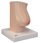
Model Of The Female Breast, Medially divided into 2 halves, held together with magnets, Healthy milk-giving breath tissue on the cut surface of the external half, Breast gland inflammation (mastitis) on the cut surface of the inner half
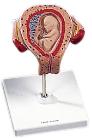
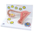
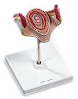
Model Stages Of Fertilisation, Dimensions: 35x21x20 cm, begin the growth into an embryo, The various stages are shown in larger-than-life model form in an ovary, fallopian tube and womb. An even more enlarged illustration of each is also printed on the base
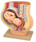
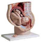
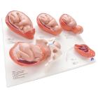
Model, Labor Stages, mounted individually bases: Fetus in womb, cervix closed, Fetus in womb, cervix open, Fetus in womb, start of head passage, Fetus in womb and pelvis, finish of head passage, Placenta in the womb, Dimensions: 40 x 31 x 13 cm, weight: 2 kg
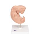
Model, Embryo, 1 month, The embryo model shows an embryo at the stage of the first month of pregnancy, Ideal for medical training and as an educational tool for pregnant women, Important anatomical structures are labeled, weight: 0.33 kg